AQUATIC EXPERIMENTAL MICROBIOLOGY PLATFORM MICROBEX
MICROBEX is structured into 3 technical facilities that cover all needs of aquatic environmental microbiology: culture, observation, enumeration, measurement of functioning and dynamics of aquatic microorganisms.
Localisation :
MARBEC
Description
-The Culture and Experimentation tray gathers equipments necessary for isolation, cultivation or breeding, and for the study of microorganisms ecophysiological processes from environmental samples or collections, to characterize their functioning in ecosystems.
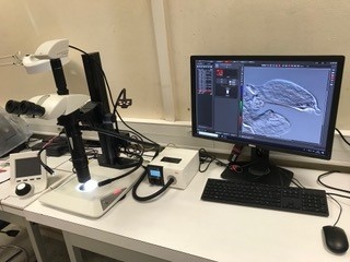
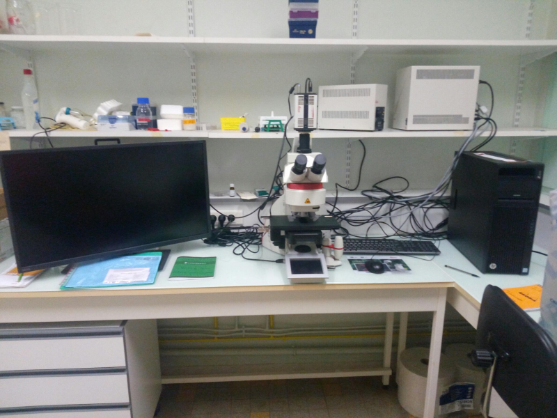
-The Microscopy-Imaging tray gathers equipments tools of preparation and observation of samples of microorganisms of 0.02µm (e.g. virus) to 6.4 cm (e.g. fish larvae). It has a space dedicated to the preparation and storage of samples fixed for microscopic or macro observations, as well as spaces (light and dark) for optimal analysis of samples. Observed microorganisms have the ability to be sorted, counted, measured, identified and images scanned with the possibility of 3D re-creation or mosaic.
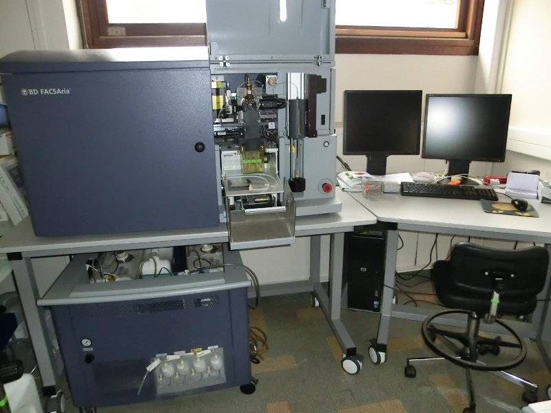
-The Flow Cytometry Flux Tray allows to count sort and physiologically characterize individual cells (30 nm to 20 µm) at high frequency (up to 50,000 cell/s), depending on their fluorescent-structure size (natural and/or induced) in response to a light stimulation (405-488-633 nm laser).
Equipment
-
- Culture and Experimentation:
The tray offers various identified spaces and dedicated equipments depending on activities and/or microorganisms specificities considering the best suited to each protocol:
・ Wetlands of land return
- Filtration stations
- Sorting stations and manipulation of various origins samples
・ Spaces for preparing culture environments
- Weight zone (accuracy scales)
- Media preparer and Petri dish distributor (Integra)
- Heated whiteers, sound-proofers, pH meters, microwave, bath
- Refrigerated cabinet and fridges for nutrients and ready-to-use sterile culture medium storing
- Tray common services support such as a laundry, a large "double door" horizontal autoclave, seawater filtration system, distilled water and MilliQ services
・ Culturing and farming areas for pathogenic (or non-pathogenic) environmental bacteria (P2-type), for phytoplankton and specifically toxic microalgae, and for zooplankton
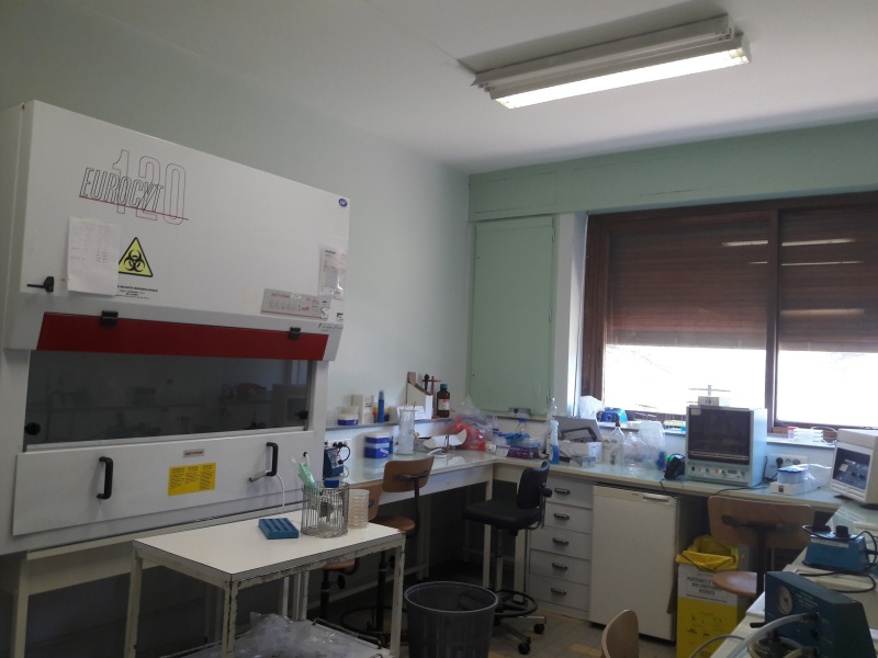
- Laminar flow hooks (PSMs) for working in a sterile environment
- Three bacteriological incubators (+ 20 / + 50 °C).
- Five thermostatically controlled incubators (- 10 / + 60 °C), with humidity and lighting control (cycle, intensity)
- air-conditioned room at 20°C with light regulation system (approx. 6 m2)
- a FLUO-STAR microplate spectrofluorimeter
- four inverted microscopes for crop control (lens with wide field depth) including one equipped with a camera
- two binocular magnifiers
- Storage equipment for welcome samples (freezers, -80 °C)
・ Spaces that offer possibilities for experimenting in microcosms and aquariums with maintaining organisms in controlled conditions
- 24 Aquariums of 30 L equipped with bubbling, pumps, and heat sinksMicroscopy-Imaging:
-A FlowCam VS-IV equipped with 3 4X, 10X and 20X lenses, a color camera and a 532 nm excitation wavelength laser for 2 emission waves (575 nm ± 15 nm and > 650 nm)
-4 epifluorescence microscopes:
IX70 (Olympus), inverted, manual, interferonal contrast, WB filters (490 nm) WG (530 nm) UV (345 nm) + color camera DFC450C, CCD
BX60 (Olympus), right, manual, phase, WB (490 nm), UV (345 nm), Chla + JENOPTIK Progress C3 cooled color camera
AX70 (Olympus), straight, semi-automated, WB(490 nm), WG (530 nm) and UV (345 nm) filters + JENOPTIK Progress MF cool monochrome camera
DM6 (Leica), right, automated, DIC, DAPI filters (345 nm), GFP, RODAMINE, Cy5, platinum XYZ + 2 cameras (1 color DMC 2900 CMOS and 1 monochrome DFC9000 GT CMOS)
-3 Light field microscopes:
AX10 Axio Imager (Zeiss), right, manual, phase contrast + SONY color camera
CK40 (Olympus), reverse, phase contrast manual
DM750 (Leica), right, manual, TP type + integrated 1CC750W digital camera
-2 binocular magnifying glass:
SZX-7 (Olympus) , x20 magnification, + camera adapter
STEMI 305 (Zeiss), type TP + camera Axiocam ERc 5s
-1 Macroscope
Z16 Apo (Leica), magnification x1 to x620, camera (Leica Axiocam 305 color) + workstation.
-1 LAS X Premium Image Analyzer (LEICA) + browser moduleCytometry
-Facs Calibur BD: 1 laser 488 nm, 4 PMT (SSC-B530-B576-B670), 1 diode FSC
-Cytoflex Beckman: 2 lasers 488-633 nm, 8 avalanche photodiode detectors (SSC-FSC-B525-B585-B690-B780-R660-R780)
-Cells Sorter Facs Aria I BD: 3 lasers 405-488-633 nm-12 PMT (B530-B576-B610-B675-B780-R660-R730-R780-V450 V530-SSC-FSC) -4 sorting paths.
Fonctionning
- Culture and experimentation:
Contact Claire Carré, the Culture and Experimentation tray manager, to assess needs and feasibility, and plan reservations for each space. It is possible to use the tray on a one-off basis or in a short to medium term (maximum 6 months). In the latter case, plan to organize yourself as soon as the projects are written to know the tray availability. A reception will then be made on the tray with a description of the good practices associated to it (plan for a DSR at least 15 days in advance). In addition, support in the implementation of manipulations and experiments can be carried out depending on the availability of the referee.
- Microscopy-Imaging:
Contact Cécile Roques, the Microscopy-Imagery tray manager. First, a project feasibility study (technical and availability) will be carried out with the project holder. Methodological tests can be carried out before a Microscopy-Imaging (UM) tray estimate can be drawn up. Registration on paper plans must be done in consultation with the manager, at least 15 days before the start of the tray. Depending on the type of project and the availability of the reference, a service delivery job may be possible.
- Flow Cytometry:
Contact Cécile Roques, the flow cytometry tray manager to assess the needs, feasibility and type of machine to be used. After quotation is issued, a purchase order must be provided. The reservation (1 to 5j max) of the schedule must be made at least 15d in advance.
Contact :
LOCATION : UM FDS bat 24, RDC
CONTACT:
- Culture and Experience: Claire Carré claire.carre@ird.fr, tel: 04 67 14 40 47
- Microscopy-Imaging: Cécile Roques cecile.roques@cnrs.fr, tel: 04 67 14 41 88
- Flow Cytometry: Cécile Roques cecile.roques@cnrs.fr, tel: 04 67 14 41 88
Documents:
Galerie photo

![]()
![]()
![]()
![]()
![]()


![]()
![]()